

Use of the best equipment, high technology
At the Stavros Tsirigotakis Model Thyroid Surgery Center, in addition to our specialization and experience, we use the highest technology every day in our operations, for the greater safety of our patients!

It is known that the expertise and experience of the thyroid surgeon are the most important factors in the safety of these operations. However, the development of technology significantly helps our effort, for even safer operations. This is why we have integrated this technology into our everyday life. Using state-of-the-art, state-of-the-art equipment in identifying and preserving the nerve and parathyroid glands, we perform the safest thyroid procedures.
You all want the best surgeon with the highest technology and that’s understandable, since it’s your most valuable asset that is YOUR HEALTH! No one should be interested in a mediocre result, especially when it has nothing to do with the cost of an operation.
Two of the rare but very unpleasant complications for patients who require thyroid surgery are damage to the nerves responsible for speech which causes hoarseness in speech and the other is damage or accidental removal of the parathyroid glands which causes hypocalcaemia (low calcium in the blood).
How is the human voice produced?
The human voice is produced by the vibrations of the vocal cords during the exhalation phase and when air passes between them.
Air during respiratory movements (inhalation, exhalation) passes through the cavity of the larynx, which is shaped like an hourglass. In the middle of the laryngeal cavity there are on either side two pairs of folds that form the false vocal cords upwards and the genuine vocal cords downwards.
During the passage of air from the lungs to the outside, the true vocal cords vibrate and produce the voice. The muscles present in the larynx control the tension of the vocal cords and the opening of the gap between them (glottis or glottis) and thus cause the different volume and frequency of the voice. Their function is controlled by the laryngeal nerves. This is why during thyroidectomy there is a possibility of causing a problem with one’s voice.
The voice acquires special characteristics in every person, in vocal resonances. These are the upper larynx region, the pharynx, the nasal cavity and the oral cavity.
In the oral cavity, with the movements of the tongue, the soft palate and the lips in relation to the teeth, the articulate speech is produced. Articulated speech is controlled by speech centers that are in the cerebral cortex and regulate the movements of these organs.
Possibility of injury to the laryngeal nerve.
Laryngeal nerve injury is one of the most feared complications of thyroid surgery. Even in experienced hands, its transient paresis occurs in 0.4-12% of cases, while its permanent paralysis is reported in up to 5-6%. Its frequency is lower (0.2-0.8%) in surgeons with a large number of patients. The nerve responsible for speech (regressive laryngeal nerve) is usually located just behind the thyroid and is a small and delicate organ that is at risk of being abused during surgery.
Nerve protection through the neurostimulator even with continuous recording – Thyroidectomy during which the patient is “talking”.

The recurrent laryngeal nerve is usually looked for by the surgeon to identify it so that it can be protected during your thyroid surgery. Identifying the nerve with the naked eye of the experienced thyroid surgeon is what has been done for many decades, however today there is high technology to help the surgeon confirm that they have correctly identified the nerve AND that the nerve is still functioning properly.

Neurostimulation equipment includes a device that sends a low-voltage signal, through a probe, to the nerve. This creates a movement, which is picked up by a special monitor via an electrode located in your breathing tube. When the surgeon locates the nerve, he touches it with a special device and then a special sound is produced which confirms that the specific organ is the nerve. In addition to this method, there is another method of continuous recording of the nerve. In this case, there is an additional device that produces a continuous sound during the operation, this sound changes when manipulations are made near the nerve, which informs the surgeon even when he has not located the nerve, that he is very close to him and he must be very careful. We can liken the operation of this device to a virtual operation in which a patient will be operated on and during the operation he will speak, so that the surgeon immediately perceives the change in the timbre of his voice. This technology helps make your thyroid surgery even safer!
Thyroid Surgeon Stavros Tsirigotakis is familiar and experienced in the use of this technology and uses it daily in all thyroid surgeries he performs.
If you need more information on this matter, please do not hesitate to contact us.
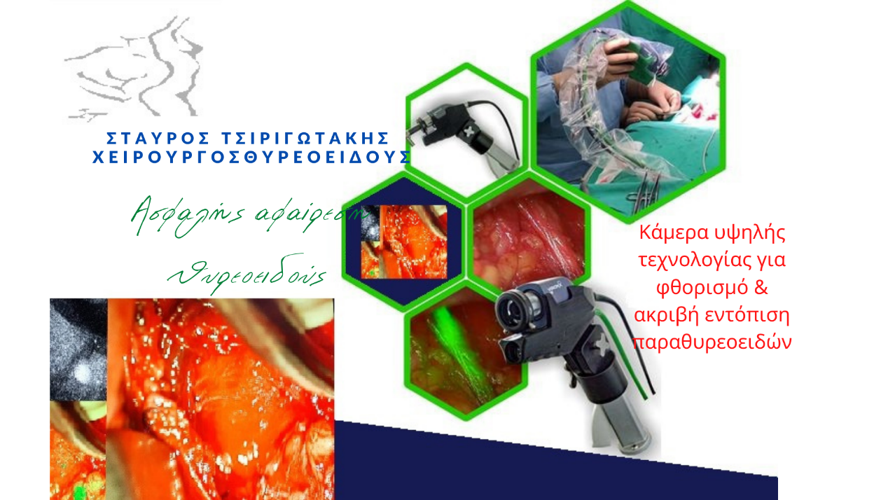
Protection of calcium and parathyroid glands during thyroidectomy, with a high-tech fluorescence camera
One potential complication that the thyroid surgeon tries to avoid is hypoparathyroidism. When performing the surgical removal of the thyroid, special care is taken to identify and preserve 4 glands usually located behind or to the side of the thyroid, called the parathyroid glands. The thyroid surgeon must identify these glands and keep them “alive” (functional).
Real-time intraoperative localization of parathyroid glands by fluorescence imaging
All thyroid surgeons report having difficulty identifying the parathyroid glands. These difficulties concern both locating the parathyroid glands and maintaining their normal function during surgery to remove the thyroid (thyroidectomy).
Calcium drop following thyroidectomy
The parathyroid glands regulate calcium levels in the body. However, their position is often not typical and sometimes it is difficult to recognize them, so that the surgeon can preserve them. This difficulty applies even to the most experienced surgeons and is unfortunately not uncommon. One of the possible complications in these operations is hypoparathyroidism and it is a serious complication mainly of total thyroidectomy. It is due to the accidental removal of the parathyroid glands or their injury. The image that the patient has in this case is not at all pleasant and sometimes it can be dangerous.
This is also the reason why in thyroid operations the patient’s calcium levels are monitored for at least two 24 hours after the operation. Hypoparathyroidism when manifested by tetany is reversed with intravenous calcium gluconate, and normal calcium levels are maintained with oral calcium tablets and vitamin D. Calcium levels are monitored weekly. Transient hypoparathyroidism usually resolves in 4 weeks, but may persist for months.
When the calcium level drops, it can cause numbness and even tetany, which is intense painful muscle contractions in the face and hands.
The symptoms are:
- muscle spasms,
- cramps with pain in arms, legs and face, or stomach,
- great weakness
- unsteadiness of walking,
- chronic hair loss
- dryness of the skin and hair,
- headache,
- concentration and memory problems,
- depression and phobias.
The use of new technology helps to avoid hypoparathyroidism in thyroid surgery

Until recently, their detection was done by identifying them with the naked eye. To overcome this existing limitation, many studies have been done on the imaging of parathyroid glands through infrared fluorescence. Fluorescence imaging of the parathyroids has been shown to be useful both in locating them and in assessing their function. This imaging technique has two parts: (I) detection through autofluorescence, using a high-tech camera, and (II) a substance (indocyanine green) is injected and through the resulting imaging, their function is checked. These innovative techniques add a significant safety advantage to performing thyroid or parathyroid surgery.
Prevention of parathyroid injury or removal in thyroidectomy
Thyroidectomy is one of the most frequently performed endocrine procedures. The prognosis for patients even with thyroid cancer is excellent. Therefore, preventing or minimizing the possibility of complications after thyroidectomy, such as injury to the nerve responsible for speech or accidental removal or hypofunction of the parathyroid glands, remains a major challenge for surgeons. The parathyroids are important organs that must be kept “alive and functional” to reduce postoperative complications in any thyroid surgery. To do this requires a very good knowledge of the anatomy, timely and accurate localization and recognition, but also very delicate manipulations and operative skill, on the part of the surgeon. Although some techniques were introduced, they were often not widely accepted. Hitherto, direct recognition with the “naked eye” and palpation, which can be acquired through experience, have been the primary tools of surgeons. Recently, biomedical engineering techniques have been studied to overcome the limitations of subjective identification and reduce the incidence of postoperative hypocalcemia (low calcium).

Basically, the first step in preserving the parathyroids and preventing the dysfunction of these organs is to correctly identify them. However, the parathyroid glands are very small organs, embedded in the paratracheal tissues, and similar in color to the surrounding tissues. In addition, their position is not always stable and can vary from patient to patient. Therefore, it may not be easy even for experienced surgeons to discover all the parathyroid glands (usually 4) with the naked eye during surgery. Thus, various methods have been studied to locate them during surgery. For hyperthyroidism, ultrasound and scintigraphy have been used with Sestamibi, with sensitivity of 69-75% and 49-70%, respectively. However, these imaging techniques are not useful in identifying a normal parathyroid. Removing part of the parathyroid gland and sending it for rapid biopsy at the time of surgery is a definitive method of histologically confirming that it is indeed a parathyroid gland, but this method can cause vascular damage and destruction of the parathyroid gland as part of it is removed. of the parathyroid. Despite the efforts that have been made, most of these methods are currently not useful and are not applied in clinical practice.

Imaging of parathyroid glands by fluorescence combined with indocyanine injection
Identification of parathyroids by fluorescence is a new field that combines medicine and optical technology. Indocyanine injection, already used elsewhere, is now applied to parathyroids. Now we are able to know in real time, during the operation, if the parathyroid we identify is also “alive” (functional).
Identification of parathyroid glands through fluorescence, with a high-tech camera

Identifying the parathyroids and distinguishing them from surrounding tissues such as the thyroid gland, fat, and connective tissue has been a challenge for surgeons. For less experienced surgeons, it is always difficult to locate and keep the parathyroid gland healthy. Even for highly experienced surgeons, inadvertent removal of one or more parathyroids has been reported to occur in up to 15% of thyroidectomy surgeries. In addition, the incidence of temporary hypoparathyroidism is up to 46% and that of permanent hypoparathyroidism is up to 6.6% after total thyroidectomy.
Fluorescent indocyanine angiography has been performed in patients undergoing thyroid surgery to visualize the vasculature of identified parathyroids. The results showed that most of the visually identified parathyroids had fluorescent indocyanine uptake. Patients who had at least one well-perfused parathyroid maintained adequate function after thyroid surgery. Prediction of parathyroid function after surgery was easier by indocyanine uptake than by visual assessment. Indocyanine angiography allows immediate assessment of vessels supplying the parathyroid that are at risk of injury during surgery. This can further help in deciding whether a parathyroid needs autotransplantation.
We have included this high technology in our everyday life, to ensure even greater safety for our patients!
If you would like to ask us something related, please contact us!